Showing posts with label quantum molecular biophysics. Show all posts

Friday, December 9, 2022

The wonders and mysteries of bioluminescence

 Members of my family have been reading Phosphorescence: On awe, wonder, and things that sustain you when the world goes dark, a personal memoir by Julia Baird.

This reminded me of how amazing and fascinating bioluminescence is, stimulating me to read more on the science side. One of the first things is to distinguish between bioluminescence, fluorescence, and phosphorescence.

Bioluminescence is chemical luminescence whereby a biomolecule emits a photon through the radiative decay of a singlet excited state that is produced by a chemical reaction. 

In contrast, fluorescence occurs when the singlet excited state is produced by the molecule absorbing a photon.

Phosphorescence occurs when a molecule emits a photon through the radiative decay of an excited triplet state, that was produced by the absorption of a photon.

Bioluminescence can occur in the dark. Fluorescence cannot as there are no photons to absorb. Phosphorescence is sometimes seen in the dark but this is because the molecule absorbs invisible UV light which produces the triplet state which has a very long radiative lifetime.

Baird gives beautiful and enchanted descriptions of seeing "phosphorescence" on her daily early morning ocean swim. She acknowledges that this is actually bioluminescence not phosphorescence. I should stress that in pointing this out I am not "unweaving the rainbow", as for literary purposes using "bioluminescent" would be clunky.


There is a useful webpage from a research group at UC Santa Barbara. They also have a detailed review article from which I took the image above.

Steven H.D. HaddockMark A. MolineJames F. Case

A much shorter review that I read this morning is

Bioluminescence in the Ocean: Origins of Biological, Chemical, and Ecological Diversity, by E.A. Widder

An article in Quanta magazine, In the Deep, Clues to How Life Makes Light by Stephanie Yin

So what is the underlying photophysics and quantum chemistry? The following review is helpful.

The Chemistry of Bioluminescence: An Analysis of Chemical Functionalities 

Isabelle Navizet, Ya-Jun Liu, Nicolas Ferré, Daniel Roca-Sanjuán, Roland Lindh

Almost all currently known chemiluminescent substrates have the peroxide bond, -O-O-, in common as a chemiluminophore. This chemical system facilitates the essential mechanism of chemiluminescence—providing a route for a thermally activated chemical ground-state reaction to produce a product in an electronically excited state. The basics of this process can be understood from studies of ... dioxetanone. [it] contains a peroxide bond, [and] fragments like the firefly luciferin system to carbon dioxide.

The squiggly line denotes the bond that is broken to produce the excited singlet state.
The figure below shows the potential energy surface that describes the dynamics leading to the emissive state. Note the presence of two conical intersections.


Much of this photophysics can be understood in terms of a "two-site Hubbard model" discussed in this classic paper that I love.

Neutral and Charged Biradicals, Zwitterions, Funnels in S1, and Proton Translocation: Their Role in Photochemistry, Photophysics, and Vision

Vlasta Bonačić-Koutecký, Jaroslav Koutecký, Josef Michl

In simple terms, all that is different in the biomolecular system is that the enzyme and the larger chromophore tune energy levels so that the energy barriers are much smaller so that the steps needed for bioluminescence become accessible at room temperature.

This highlights two fundamental things. 

Chemistry is local. This is relevant to understanding Wannier orbitals in solid state physics, to hydrogen bonding, and how protein structure aids function.

"Biochemistry is the search for the chemistry that works" [in water at room temperature].

Thursday, November 25, 2021

Role of quantum nuclear motion in biomolecular systems

 Total I am giving a talk, "Effect of quantum nuclear motion on hydrogen bonds in complex molecular materials" at Light-matter Interactions from scratch: Theory and Experiments at the Border with Biology 

Here are the slides

The talk provides a concrete example of the tutorial on constructing simple model Hamiltonians for complex materials that I give before the talk. It relates to the bio theme of the meeting through work on isotopic fractionation in proteins and the recent paper below. It makes use of the simple model that I talk about.

Unusual Spectroscopic and Electric Field Sensitivity of Chromophores with Short Hydrogen Bonds: GFP and PYP as Model Systems

Chi-Yun Lin and Steven G. Boxer

Tuesday, November 23, 2021

Tutorial on modelling quantum dynamics in biomolecules

This week I am giving two (virtual) talks at a meeting

Light-matter Interactions from scratch: Theory and Experiments at the Border with Biology 

supported by the ICTP (International Center for Theoretical Physics) in Trieste.

In the ICTP tradition, one talk is a tutorial and the second talk is about my research.

Here are the slides for the tutorial on Effective Model Hamiltonians for Quantum Dynamics in Complex Molecular Materials. Feedback is welcome.

The research talk is about hydrogen bonding. I will post slides for that later.


Thursday, March 5, 2020

The quantum physics of life in red and green

Life is truly amazing!
Life is beautiful!
...and it involves quantum many-body physics...

There is a beautiful (short) review
Heme: From quantum spin crossover to oxygen manager of life 
Kasper Kepp

The article involves a plethora of topics that I have discussed before on this blog. I have included relevant links.

Kepp starts with the unique (chemically fine-tuned) properties of both iron and porphyrin that enable them to play a central role in two of the most important processes in life: respiration and photosynthesis. He has a beautiful paragraph (perhaps in the style of Roald Hoffmann):
Such ligand-field transitions of iron in porphyrin were familiar to our ancestors as the characteristic red color of blood that largely defines the human psychological and cultural connotations of the color representing courage, war, danger, and suffering. 
Incidentally, pi-pi* transitions within the porphyrin-derived chlorophylls are also responsible for the green color of plants, associated with nature, life and hope, so the reader may perhaps agree that porphyrin has had vast (but alas! rarely appreciated) cultural consequences.
The oxygen molecule is a spin triplet.
Iron(II) porphyrin is in a triplet spin state (S=1). The Fe(II) is a d6 configuration in a D_4h crystal field.
When they bind together the ground state is a spin-singlet.

There are two fundamental quantum chemistry questions that are discussed.

1. What is the electronic structure (many-body wave function) of the ground state for oxygen bound to heme?

2. What is the mechanism for the ``spin-forbidden'' transition of the oxygen binding?

The first question has a long history. Like almost anything important and profound in quantum chemistry it goes back to Linus Pauling! In 1936 Pauling and Coryell argued that the ground state is
essentially a neutral O=O binding with two of its electrons to iron to produce a formally iron(II) if both the bonding electrons were confined to O2, corre- sponding to the non-bonding limit of neutral parts, but a formally iron(I) if the Fe–O bond were to be considered covalent. 
In 1960, McClure suggested a valence-bond formulation based on triplet–triplet coupling, which is appealing by the low promotion energies required to access these states, rather than the singlet states. In 1964, Weiss suggested, based on analogy to chemical reactions in aqueous solution, that the true ferrous hemeO2 adduct was mainly of the superoxo-iron(III) type caused by ‘‘electron transfer” from iron to O2. 
Goddard and Olafson suggested an ozone model of the adduct in 1975 which emphasized the four-electron three-center bond with maintained triplet state of dioxygen as in the McClure model with less electronic reorganization to explain the reversible binding. 
In 1977, Pauling maintained his original view again, the same year that Huynh, Case, and Karplus did a first attempt to bridge these views by performing early quantum chemical calculations that diplomatically emphasized the importance of both Weiss and Pauling resonance forms. 
However, interpretation depends on model language, orbital localization, and transformation between valence bond and orbital formalisms:  
In terms of molecular orbital theory, the wave function was a multi-configurational state dominated by the Pauling configuration; however, if one uses valence bond theory considerations, it can be interpreted as having large Weiss character. Thus, the multi-configurational state produced from CASPT2 is interpreted differently by different models. This partly explains why the trenches were so deeply dug during the exchange between Pauling, Goddard, McClure, and Weiss; all were right, and all were wrong. 
This is just another example of unnecessary conflicts about valence bond vs. molecular orbital (VB vs. MO). 
In terms of valence structures, the ground state was summarized by Shaik and Chen as having contributions from both Weiss, Pauling, and McClure forms, the first .. dominating. 
Ironically DFT ends up providing a useful language after all! 
The charge assignments to O2 are very dependent on calculation scheme, and both the orbitals, valence structures, and atomic charges that defined the Weiss-Pauling debate are non-observable. In contrast, the electron density is observable as are the geometries and spectroscopic data...
Molecular orbitals are not physical observables but calculational constructs. MO's don't exist.

In different words, one can take a many-body wave-function and make a linear unitary transformation of the molecular orbitals. The Slater determinants do not change. [The value of a determinant is invariant to a change of basis.]

Now. Question 2.
What is the mechanism for the ``spin-forbidden'' transition of the oxygen binding?

Kepp talks about spin-orbit coupling and the fact that it is small for oxygen, motivating a discussion of a "broad crossing mechanism".  However, I am not sure this is relevant. I don't see the binding as necessarily spin forbidden. As the oxygen approaches the heme the two triplet states can mix to form a total spin singlet.
This is analogous to bringing two hydrogen atoms (each of which is spin 1/2) together to form a hydrogen molecule (which is spin zero). A multi-configurational wavefunction has no problem with this. But DFT-based approximations, which use a single determinant cannot describe this smooth crossover.

Other things of particular interest to me that are discussed in the review include the central role of back bonding and the success of the TPSSh functional in DFT calculations for organometallics.

Unfortunately, the review does not mention recent work by Weber et al, applying DMFT to the problem of oxygen binding to haemoglobin.

Friday, May 5, 2017

Talk on "crackpot" theories

At UQ there is a great student physics club, PAIN. Today they are having a session on "crackpot" theories in science. Rather than picking on sincere but misguided amateurs I thought I would have a go at "mainstream" scientists who should know better. Here are my slides on quantum biology.

A more detailed and serious talk is a colloquium that I gave six years ago. I regret that the skepticism I expressed then seems to have been justified.

Postscript.
I really enjoyed this session with the students. Several gave interesting and stimulating talks, covering topics such as flat earth, last thursdayism, and The Final Theory of gravity [objects don't fall to the earth but rather the earth rises up to them...]. There were good discussions about falsifiability, Occam's razor, Newton's flaming laser sword, ...
There was an interesting mixture of history, philosophy, humour, and real physics.

I always find to encouraging to encounter students who are so excited about physics that they want to do something like this on a friday night.

Wednesday, November 30, 2016

Photosynthesis is incoherent

Beginning in 2007 luxury journals published some experimental papers making claims that quantum coherence was essential to photosynthesis. This was followed by a lot of theoretical papers claiming support. I was skeptical about these claims and in the first few years of this blog wrote several posts highlighting problems with the experiments, theory, interpretation, and hype.

Here is a recent paper that repeats one of the first experiments.

Nature does not rely on long-lived electronic quantum coherence for photosynthetic energy transfer Hong-Guang Duan, Valentyn I. Prokhorenko, Richard Cogdell, Khuram Ashraf, Amy L. Stevens, Michael Thorwart, R. J. Dwayne Miller
During the first steps of photosynthesis, the energy of impinging solar photons is transformed into electronic excitation energy of the light-harvesting biomolecular complexes. The subsequent energy transfer to the reaction center is understood in terms of exciton quasiparticles which move on a grid of biomolecular sites on typical time scales less than 100 femtoseconds (fs). Since the early days of quantum mechanics, this energy transfer is described as an incoherent Forster hopping with classical site occupation probabilities, but with quantum mechanically determined rate constants. This orthodox picture has been challenged by ultrafast optical spectroscopy experiments with the Fenna-Matthews-Olson protein in which interference oscillatory signals up to 1.5 picoseconds were reported and interpreted as direct evidence of exceptionally long-lived electronic quantum coherence. Here, we show that the optical 2D photon echo spectra of this complex at ambient temperature in aqueous solution do not provide evidence of any long-lived electronic quantum coherence, but confirm the orthodox view of rapidly decaying electronic quantum coherence on a time scale of 60 fs. Our results give no hint that electronic quantum coherence plays any biofunctional role in real photoactive biomolecular complexes. Since this natural energy transfer complex is rather small and has a structurally well defined protein with the distances between bacteriochlorophylls being comparable to other light-harvesting complexes, we anticipate that this finding is general and directly applies to even larger photoactive biomolecular complexes.
I do not find the 60 fsec timescale surprising. In 2008, Joel Gilmore and I published a review of experiment and theory on a wide range of biomolecules (in a warm wet environment) that suggested that tens of femtoseconds is the relevant time scale for decoherence.

I found the following section of the paper (page 7) interesting and troubling.
The results shown in Figs. 3 (a) and (b) prove that any electronic coherence vanishes within a dephasing time window of 60 fs. It is important to emphasize that the dephasing time determined like this is consistent with the dephasing time of τhom = 60 fs independently derived from the experiment (see above). It is important to realize that this cross-check constitutes the simplest and most direct test for the electronic dephasing time in 2D spectra. In fact, the only unique observable in 2D pho- ton echo spectroscopy is the homogeneous lineshape. The use of rephasing processes in echo spectroscopies removes the inhomogeneous broadening and this can be directly inferred by the projection of the spectrum on the antidiagonal that shows the correlation between the excitation and probe fields. This check of self-consistency has not been made earlier and is in complete contradiction to the assertion made in earlier works. Moreover, our direct observation of the homogeneous line width is in agreement with independent FMO data of Ref. 12. This study finds an ∼ 100 cm−1 homogeneous line width estimated from the low-temperature data taken at 77 K, which corresponds to an electronic coherence time of ∼ 110 fs, in line with our result given the difference in temperature. In fact, if any long lived electronic coherences were operating on the 1 ps timescale as claimed previously (1), the antidiagonal line width would have to be on the order of 10 cm−1, and would appear as an extremely sharp ridge in the 2D inhomogeneously broadened spectrum (see Supplementary Materials). The lack of this feature conspicuously points to the misassignment of the long lived features to long lived electronic coherences where as now established in the present work is due to weak vibrational coherences. The frequencies of these oscillations, their lifetimes, and amplitudes all match those expected for molecular modes (41, 42) and not long-lived electronic coherences.

Wednesday, November 23, 2016

How I got a Wikipedia page

It has dubious origins.

Some people are very impressed that I have a Wikipedia page.
I find this a bit embarrassing because there are many scientists, more distinguished than I, who do not have pages.
When people tell me how impressed they are I tell them the story.

Almost ten years ago some enthusiasts of "quantum biology" invited me to contribute a chapter to a book on the subject. The chapter I wrote, together with two students, was different from most of the other chapters because we focussed on realistic models and estimates for quantum decoherence in biomolecules. (Some of the material is here.) This leads one to be very skeptical about the whole notion that quantum coherence can play a significant role in biomolecular function, let alone biological processes. Most other authors are true believers.

I believe that to promote the book one of the editors had one of his Ph.D. students [who appeared to also do a some of the grunt work of the book editing] create a Wikipedia page for the book and for all of the senior authors. These pages emphasised the contribution to the book and the connection to quantum biology.

The "history" of my page states it was created by an account that
An editor has expressed a concern that this account may be a sock puppet of Bunzil (talk · contribs · logs).
I have since edited my page to remove links and references to the book since it is not something I want to be defined by.

An aside. Today I updated the page because when giving talks I got tired of sometimes being introduced based on outdated information on the page.

Hardly, a distinguished history....


The xkcd cartoon is from here.

Monday, June 20, 2016

A nice text on spectroscopy of biomolecules

Bill Parson kindly gave me a copy of the new edition of his book, Modern Optical SpectroscopyWith Exercises and Examples from Biophysics and Biochemistry
It is a excellent book that covers a range of topics that are of increasing importance and interest to a range of people.
I am not sure I am aware of any other books with similar scope.

Two particular audiences will benefit from engaging with the material.

1. Biochemists and biophysics who have a weak background in quantum theory and need to understand how it underpins many spectroscopic tools that are now widely used to describe and understand biomolecules.

2. Quantum physicists who are interested in the relevance (and irrelevance!) of quantum theory to biomolecular systems. For some it could be a reality check of the complexities and subtleties involved and the long and rich history associated with the subject.

I highly recommend it. I have learnt a lot from it, some it quite basic stuff I should have known.

Monday, July 27, 2015

Quantum biology smells bad

I am skeptical of the grand and speculative claims of "quantum biology". 
There is a nice paper in PNAS which systematically considers the specific claim that smell is based on sensing the vibrational frequencies of particular molecules, and rebuts it from both theoretical and experimental points of view.

Implausibility of the vibrational theory of olfaction
Eric Block, Seogjoo Jang, Hiroaki Matsunami, Sivakumar Sekharan, Bérénice Dethier, Mehmed Z. Ertem, Sivaji Gundala, Yi Pan, Shengju Li, Zhen Li, Stephene N. Lodge, Mehmet Ozbil, Huihong Jiang, Sonia F. Penalba, Victor S. Batista, and Hanyi Zhuang.

I thank Suggy Jang for bringing the paper to my attention.

Monday, July 6, 2015

Genetically engineering short hydrogen bonds in a fluorescent protein

There is a very nice article in the new journal, ACS Central Science
Short Hydrogen Bonds and Proton Delocalization in Green Fluorescent Protein (GFP) 
Luke M. Oltrogge and Steven G. Boxer

This is an impressive piece of work spanning from molecular biology to chemistry to quantum physics.
There is also a commentary on the paper by Judith Klinman, placing it in the context of the controversial issue of low-barrier hydrogen bonds in enzymes.

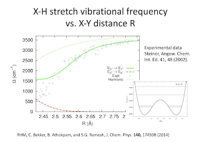
An extensive study was made of mutants of the Green Fluorescent Protein with a short hydrogen bond between the chromophore and the amino acid Asp148. The donor-acceptor bond length estimated from X-ray structures was 2.4 +/- 0.2 Angstroms. This is in the range of low-barrier H-bonds.

What is particularly new here is that through ingenious molecular biology techniques [nonsense suppression] the acidity [pK_a = measure of tendency to give up protons] of the chromophore was systematically varied by 3.5 units through halogen substitutions.


This range covers the pK_a matching required for strongest H-bonds, as discussed in this earlier post. The experimental results were compared to calculations based on a one-dimensional proton transfer potential based on a diabatic state model I have advocated. It was very satisfying for me to see this simple model being used by experimentalists.

To me what is most striking about the paper is the UV absorption spectra below. It is very different from what one normally sees in GFP spectra.
There are generally two absorption bands, denoted A and B, associated with GFP. The A-state and B-state are identified with the neutral chromophore and anionic [i.e. deprotonated] chromophore, respectively. The corresponding spectra are similar to the black and grey curves shown above. The green spectrum above is for the Cl1Y substituted chromophore, which is close to pK_a matching, and is rather broad and intermediate between the A-state and B-state spectra. This is arguably because the proton is delocalised between the chromophore and neighbouring Asp amino acid.

The authors also substituted protons (H) with deuterium (D) to see the extent of quantum nuclear effects. These are normally very small in GFP. However, here they are noticeable.
The measured isotopic fractionation factors Phi (deduced from analysis of the UV absorption spectra) were in the range 0.54 - 0.9, taking a minimum value for pK_a matching. This observation and a value of Phi=0.54 for R=2.4 +/- 0.2 Angstroms are consistent with a recent theoretical analysis.

There is one point where I disagree with the theoretical analysis of the authors. I am confused that they average over the vibrational eigenstates to get an electronic absorption spectrum. This seems to me this goes against the Franck-Condon principle.  If one followed this same procedure for other molecules the UV spectra would all be much broader than they are, particularly in gas phase.
It is not clear to me how one should proceed in this situation where the proton is quite delocalised and the absorption spectra is significantly different for protonated and de-protonated chromophores. There may be significant Herzberg-Teller effects. One way forward to could be to combine the two-diabatic state H-bond model with a two-state resonance model for the chromophore, such as those advocated by Seth Olsen and I, and then do a full non-adiabatic treatment of the model.

I thank Luke Oltrogge and Seth Olsen for helpful discussions about this work.

Tuesday, April 21, 2015

Calibrating a ruler for hydrogen bond lengths

I have just finished a paper with Bijyalaxmi Athokpam and Sai  Ramesh,
Isotopic fractionation in proteins as a measure of hydrogen bond length

If a deuterated molecule containing strong intramolecular hydrogen bonds is placed in a hydrogenated solvent it may preferentially exchange deuterium for hydrogen. This preference is due to the difference between the vibrational zero-point energy for hydrogen and deuterium.  It is found that the associated fractionation factor $\Phi$  is correlated with the strength of the intramolecular hydrogen bonds. This correlation has been used to determine the length of the H-bonds (donor-acceptor separation) in a diverse range of enzymes and has been argued to support the existence of short low-barrier H-bonds.

Starting with a potential energy surface based on a simple diabatic state model for H-bonds we calculate $\Phi$ as a function of the proton donor-acceptor distance $R$.  For numerical results, we use a parameterization of the model for symmetric O-H.... O bonds.  We consider the relative contributions of the O-H stretch vibration, O-H bend vibrations (both in plane and out of plane), tunnelling splitting effects at finite temperature, and the secondary geometric isotope effect. We
compare our total $\Phi$ as a function of $R$ with NMR experimental results for enzymes, and in particular with an empirical parametrisation $\Phi(R)$, used previously to determine bond lengths.

I welcome any comments or suggestions.

Wednesday, December 10, 2014

Strong non-adiabatic effects in a prototype chemical system

This post concerns what may be the fast known internal conversion process in a chemical system, non-radiative decay times in the range of 3-8 femtoseconds. Internal conversion is the process whereby in a molecule there is a non-radiative transition between electronic excited states (without change in spin quantum number). This is by definition a break-down of the Born-Oppenheimer approximation.

Much is rightly made of the fascinating and important fact that excited states of DNA and RNA undergo "ultra-fast" non-radiative decay to their electronic ground state. This photo-stability is important to avoid mutations and protect genetic information. Conical intersections are key. The time scale for comparison is the order of a picosecond.

The figure below is taken from

It shows the wavelength dependence of the intensity of emission from a 3d (Rydberg) excited state.

There are several things that are noteworthy about the experimental data, given that this is a gas phase spectra.

1. The large width of the spectra. In energy units this is of the order of an eV. Gas phase spectra for electronic transitions in typical molecules are usually extremely sharp (See here for a typical example). 

2. The two peaks, suggesting the presence of two electronic transitions.

3. The strong isotope effects. For strictly electronic transitions between adiabatic states, there should be no dependence on the nuclear masses. This suggests strong vibronic and quantum nuclear effects.

So what is going on?
The key physics is that of the Jahn-Teller effect, conical intersections, and non-adiabatic effects.
For H3 there is geometry of an equilateral triangle which has C3 symmetry. There are then two degenerate electronic ground states with E symmetry, and experience E x epsilon Jahn-Teller effect leading to the two adiabatic potential energy surfaces shown below. They touch at a conical intersection. The two peaks in the spectra above correspond to transitions to these two different surfaces.

Non-adiabatic coupling leads to rapid transitions between the surfaces leading to the ultra-ultra-fast internal conversion and the very broad spectra. This is calculated in the paper, leading to the theoretical curves shown in the top figure.


More recently, Susanta and some of his students, have considered the relative importance of (off-diagonal) non-adiabatic effects, the geometric phase [associated with the conical intersection], and Born-Huang (diagonal) corrections to explaining the spectra.
They find that the first has by far the most dominant effect. The latter two have very small effects that look like they will be difficult to disentangle from experiment. I discussed the elusiveness of experimental signatures of the geometric phase in an earlier post.
  
I thank Susanta Mahapatra for explaining this nice work to me, on my recent visit to his group.

Friday, September 5, 2014

The challenge of coupled electron-proton transfer

There is a nice helpful review
Biochemistry and Theory of Proton-Coupled Electron Transfer 
Agostino Migliore, Nicholas F. Polizzi, Michael J. Therien, and David N. Beratan

Here are a few of the (basic) things I got out of reading it (albeit on a long plane flight a while ago).

There are a diverse range of biomolecules where coupled electron-proton transfer plays a key role in their function. The electron transfer (ET) and proton transfer (PT) are usually spatially separated. [See blue and red arrows below].

There are fundamental questions about whether the transfer is concerted or sequential, adiabatic or non-adiabatic, and how important the protein environment (polar solvent)  is.

Often short hydrogen bonds are involved and so the nuclear degrees of freedom need to be treated quantum mechanically, in order to take into account tunnelling and/or zero-point motion.

Diabatic states are key to understanding and theoretical model development.

Although there are some "schematic" theories, they involve some debatable approximations (e.g. Fermi's golden rule), and so there is much to be done, even at the level of minimal model Hamiltonians.

Wednesday, June 25, 2014

Condensed phase dynamics in Telluride

Last night I was stranded at Denver airport en route to the bi-annual Condensed phase dynamics meeting at the Telluride Science Research Center.  This is the third time I have been to this wonderful meeting. Getting there can be a real hassle. But, then you look at the scenery and enjoy the science and it seems worth it.


Unfortunately, due to the travel delays I missed the first two talks, by Joe Subotnik and Nandini Ananth.

Dominika Zgid gave a chemist's perspective on "How to make dynamical mean theory quantitative". Some of her work was discussed in a my last post. Today she mostly discussed a generalisation of iterative perturbation theory as an "impurity solver" for DMFT problems with multiple orbitals. See this preprint.

Peter Rossky discussed quantum chemical simulations of exciton dynamics in conjugated polymers.

This was motivated by an experiment reported in Science that claimed evidence for quantum coherent transport of excitons along a polymer chain at room temperature. Several oscillations were seen in the fluorescence polarisation anisotropy  as it decays in about a picosecond. These oscillations were identified with quantum inference [Rabi oscillations] between different exciton states delocalised over the polymer chain.

It turns out the experimental results have a much more mundane explanation.
The simulations of Adam Willard and Rossky are of classical dynamics on the adiabatic excited state potential energy surface calculated from a parameterised PPP [Pariser-Parr-Pople] model [basically a Hubbard model with long-range Coulomb interactions. They see oscillations similar to those in the experiment and can identified simply with classical nuclear motion associated with the polymer backbone stretching [phonons] in response to photo-excitation.

Much-hyped experiments claiming to show quantum coherence in photosynthetic complexes, probably also have a similar classical explanation in terms of nuclear dynamics rather than electronic coherences. A concrete interpretation in terms of vibrational coherences is in this PNAS paper. My skepticism of these "quantum biology" experiments has been expressed in many earlier posts.

Hopefully, tomorrow I will blog about talks from Eran Rabani, Todd Martinez, and Dvira Segal.

Wednesday, March 26, 2014

Desperately seeking low-barrier hydrogen bonds

There is an interesting JACS article
Are There Really Low-Barrier Hydrogen Bonds in Proteins? The Case of Photoactive Yellow Protein
Marc Nadal-Ferret, Ricard Gelabert, Miquel Moreno, and José M. Lluch

Low-barrier hydrogen bonds are characterised by an energy barrier to proton transfer that is comparable to the vibrational zero-point energy. As a consequence the proton is delocalised between the donor and the acceptor atoms.

Previously I posted about the general issue of whether these bonds exist in proteins, and more importantly whether they have a functional role. Before I started working on H-bonds I wrote a post about new experimental studies claiming that the photoactive yellow protein has a low barrier H-bond (LBHB). The relevant geometry and the two relevant H-bonds [2.52 and 2.56 Angstroms] are shown below.
The key issue this JACS paper addresses is
in several very recent papers, Saito and Ishikita (33-35) have claimed that …  the chemical properties of the pCA···Glu46 bond can be simply explained as a conventional hydrogen bond, without invoking the LBHB concept. In particular,(33) they have carried out quantum mechanical/molecular mechanical (QM/MM) calculations to reproduce the two short hydrogen bond distances of the crystal structure, obtaining 2.57 and 2.50 Å for pCA···Glu46 and pCA···Tyr42, respectively, but they have not found any minimum energy structure with the proton near the central region of the hydrogen bonds. In both cases, the electronic structure calculations lead to energy minima with the two protons clearly belonging to the Glu46 or Tyr42 moieties, respectively.
However, as the authors stress, this earlier work treats the proton classically. The JACS takes into account quantum effects of the proton motion.

They find a low-dimensional ground state potential energy surface using several QM/MM methods [most DFT with the CAM-B3LYP functional] and then find the low-lying vibrational eigenstates, for both protons and deuterium. They conclude
our work supports the dual result that, in the solid (crystal) phase, PYP presents an LBHB in the pCA···Glu46 hydrogen bond, whereas in solution this strong interaction is gone and shows characteristics of a “normal” hydrogen bond, much in line with what was found for many simpler systems by Perrin et al. (26, 27) Then our results support the first direct experimental demonstration of the formation of an LBHB in a protein.
I am wondering how robust these results are. There are some extreme subtleties.

First, the results for the potential energy surface for proton transfer may depend significantly on the quantum chemical method  that is used and on the method for dividing the QM/MM region.

Second, following some of the results in my recent preprint, when the donor-acceptor distance R is about 2.5 Angstroms (A) quantum effects become very significant leading to
  • an increase in the average value of R from the minimum of the classical potential by about 0.1 A due to zero-point energy of the proton.
  • a difference of about 0.05 A between hydrogen and deuterium. This is significant because it means the distances determined from neutron scattering on deuterated crystals will be different from the native protein with protons.
  • a significant change in the proton transfer potential [particularly the size of the energy barrier] as R changes by amounts as small as 0.05 A.
Due to all of the above I think that it is going to be difficult to make definitive conclusions about this fascinating and important issue.

Tuesday, March 18, 2014

Nanoscale Schrodinger kittens and double proton transfer

I think double proton transfer can be pretty amazing. The picture below shows two possible quantum states of a porphycene molecule. Note two things.

First, the two states differ by the location of the two hydrogen atoms [protons].
Second, the location of eight of the double bonds is different.


At low temperatures the ground state of the molecule is a linear superposition of the two states.
The definitive signature of this is the tunnel splitting of the vibrational states, as shown above.
This is seen experimentally, as reported here.
At higher temperatures does not see a splitting and there is a temperature activated conversion between the two tautomers.

The ground state can be written as a superposition of two Born-Oppenheimer states [products of nuclear and electronic wave functions]

 Psi = |L>|A> + |R>|B>

where |L> and |R> are the two nuclear states and |A> and |B> the two electronic states. 
These are approximately orthogonal to each other.

What is impressive about this?
Each electronic state involves about 28 valence electrons. Roughly each double bond can be described by a valence bond state consistent of a pair of electrons in a maximally entangled singlet state.

This is not quite a Schrodinger cat state. But it is a nanoscale kitten!

How is such a state possible? The key is that the two electronic states are very strongly coupled. They have a Hamiltonian matrix element of order of electron volts [10,000 cm^-1] . Yet the tunnel splittings are only a few cm^-1 due to the small overlap of nuclear states.
Hence, these superposition states are very fragile and will be easily destroyed at a few kelvin and/or any sort of polar solvent. So don't even start thinking about quantum biology!

Wednesday, February 5, 2014

Quantum fluctuations protect your genetic code


Yesterday I read an interesting paper
Enol Tautomers of Watson−Crick Base Pair Models Are Metastable Because of Nuclear Quantum Effects
Alejandro Pérez, Mark Tuckerman, Harold Hjalmarson, and Anatole von Lilienfeld

A key to the double helix structure of DNA and its ability to provide reliable stable storage of genetic information is hydrogen bonding between base pairs [G-C and A-T].
However, it is possible to switch around the positions of the protons on each of the base pairs, producing different tautomers of T, A, C, and G]. 
This is an example of double proton transfer.

This could lead to problems with correctly storing genetic information. 
An important question concerns just how rare this is. For example, what is the free energy of these tautomers relative to the Watson-Crick ones?
Over the past two decades a number of classical molecular dynamics simulations, using potentials derived from quantum chemistry suggested that the tautomers of DNA could be a problem.


Well, the paper above actually shows/argues [based on ab initio path integral molecular dynamics] that the quantum motion of the protons destabilises the mutant tautomers.

Thursday, August 22, 2013

Defining non-trivial quantum effects in chemical dynamics

Bill Miller has a very nice Perspective: Quantum or Classical coherence? in the Journal of Chemical Physics. I thank him for explaining some of it to me today.

He clearly defines what he considers to be a truly quantum effect in chemical dynamics.
It is particularly interesting because by his definition Rabi oscillations are not quantum. They are just like two coupled classical harmonic oscillators.

He starts with a Feynman path integral representation of some time-dependent correlation function and considers the semi-classical (SC) limit. The correlation function can be written as an initial value representation (IVR). If one linearises the paths (LSC) one obtains classical Wigner functions and one cannot capture quantum interference effects [e.g. double slit interference which involves paths with more than infinitesimal separation].

Tao and Miller considered the semi-classical path-integral representation of the spin-boson model. They use the Meyer-Miller-Stock-Thoss representation to map the "spin" [two-level system] to a pair of harmonic oscillators [for condensed matter physicists these are Schwinger bosons]. This allows a semi-classical treatment of the two-level system. Then the Rabi oscillations are just like transfer of energy backwards and forwards between two coupled classical harmonic oscillators. This leads to the figure below.
The "Present model" refers to the LSC-IVR treatment, i..e there is no quantum interference.

Ishizaki-Fleming refers to a much more sophisticated "quantum" treatment working towards describing the much-hyped [and probably mistaken] "quantum coherence" of exciton transfer in photosynthetic systems.

The article also contains several other concrete examples: some with and some without quantum coherence. A Tully non-adiabatic problem is particularly interesting because the nuclei exhibit quantum coherence but the electrons don't.

Wednesday, July 31, 2013

How to (not) break into a new field

Over the years I have moved into new established research areas with mixed success.
Sometimes this has been a move from one sub-field of condensed matter theory to another. Other times it has been to try and cross disciplines, e.g. into theoretical chemistry.
I have also watched with interest as others have tried to break into communities I have been a part of.

Here is my list of suggestions as to things that may increase your chances of success.

1. Listen.
What do the well established experts in the field say? What are they working on? What do they think are the important questions? What are the key concepts and landmarks in the field?
Bear in mind the values and culture may be quite different from your own field.
Note: this is the economist Paul Krugman's first research tip.

2. Be humble.
Most fields have a long history and have been pioneered by some very smart and hard working people. That doesn't mean that the field isn't populated by some mediocre people or bad ideas or bad results or that you have nothing to contribute. But, don't presume that you actually understand the field or that your new "idea" or "contribution" is going to be well received. Treating people in the field with disdain and/or making bold unsubstantiated claims will increase resistance to your ideas. Don't be the scientist who cried "Breakthrough!"

3. Be patient.
Don't expect people to instantly understand what you are on about or how important your contribution is. It may take years or decades to be accepted.

4. Make personal connections.
Go to conferences. Talk to people. Slowly and carefully explain what you are doing. Learn from them.

Who is your real audience?
Are you just trying to impress your department chair, a funding agency, or your home discipline, by claiming that what you are doing is relevant to other research areas? Or are you actually trying to make a real contribution to a different community?

If you want to see a case of physicists not being well received in biology read Aaaargh! Physicists! Again! by PZ Myers about a new theory of cancer proposed by Paul Davies and Charles Lineweaver.
An article by Davies about his theory is the cover story of the latest issue of Physics World.

I welcome suggestions and anecdotes.

Saturday, July 20, 2013

Are large atomistic quantum dynamical simulations falsifiable?

There are a whole range of dynamical processes found in biomolecular systems that one would like to simulate and understand. Examples include:
  • charge separation at the photosynthetic reaction centre
  • proton transfer in an enzyme
  • photo-isomerisation of a fluorescent protein
  • exciton migration in photosynthetic systems.
These are particularly interesting and challenging because they lie at the quantum-classical boundary. There is a subtle interaction between a quantum subsystem [e.g. the ground and excited electronic states of a chromophore] and the environment [the surrounding protein and water] whose dynamics are largely classical.


[image from here]

Due to recent advances in computational power and new algorithms [some based on conceptual advances] it is now possible to perform simulations with considerable atomistic detail. This is an example of "multi-scale" modeling.

However, it is important to bear in mind the many ingredients and many approximations employed, at each level of the simulation. These can include

  • uncertainty in the actual protein structure
  • the force fields used for the classical dynamics of the surrounding water 
  • the atomic basis sets used in "on the fly" quantum chemistry calculations 
  • density functionals used [if DFT is a component] 
  • surface hopping for non-adiabatic dynamics [e.g. non-radiative decay of an excited state].
These approximations can be quite severe. For example, a widely used water force field used in biomolecular simulations, TIP3P, predicts that water freezes as -130 degrees Centigrade!

Then there is the whole question of how one decides on how to divide the quantum and classical parts of the simulation.


If there are 10 approximations involving errors of 20 per cent each, what is the likely error in the whole simulation? Obviously, it is actually much more complicated than this. Will the errors be additive or independent of one another? Or is one hoping for some sort of cancellation of errors?

What is one really hoping to achieve with these simulations?
In what sense are they falsifiable? 
When they disagree with experiment what does one conclude?

An earlier post highlighted the critical assessment  of one expert, Daan Frenkel of the role of classical simulations. I hope we will see a similar assessment of quantum simulations in molecular biophysics.

The role of superconductivity in development of the Standard Model

In 1986, Steven Weinberg published an article,  Superconductivity for Particular Theorists , in which he stated "No one did more than N...